

The Scientific Laboratories are equipped with state-of-the-art instrumentation, used for conducting analytical campaigns that support archaeological and art historical scholarship, conservation treatments, and educational activities related to the University of Turin’s Master’s Degree. This equipment is also employed to implement environmental monitoring programs and to develop research projects at the national and international level.
The available techniques include both non-invasive and micro-invasive methods, such as 2D and 3D imaging, optical and electron microscopy, elemental and molecular spectroscopies, biological investigations, and environmental monitoring systems.
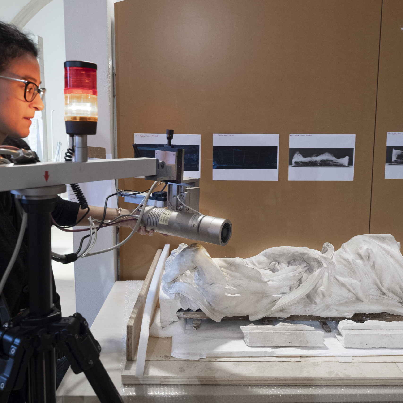
Additionally, the Laboratories house a unique radio-tomographic system specifically designed for examining large-scale artifacts. This technology was assembled within the neu-ART research project (2010-2013), funded by the Piedmont Region and developed in collaboration with the University of Turin’s Department of Physics and with the Turin branch of the National Institute for Nuclear Physics (INFN).
INSTRUMENTATION AND ANALYTICAL TECHNIQUES
Below is a list of the instruments and analytical techniques available at the Scientific Laboratories:
Photographic and Imaging Investigations
- Visible diffuse and raking light photography
- Visible light transillumination photography
- Infrared light transillumination photography
- Infrared reflectography (IRR, 850-1000 nm) with false color processing
- Ultraviolet reflectography (UVR) with false color processing
- Ultraviolet-induced visible fluorescence (UVF)
- Visible-induced infrared luminescence (VIL)
- Reflectance transformation imaging (RTI)
- Photogrammetry
- 3D scans with laser triangulation scanner
- 3D scans with a time-of-flight laser scanner
- Digital radiography (up to 4 m x 3 m)
- Computed tomography (up to 2.5 m height x 2 m base diameter)
Non-Invasive Spot Analysis
- Portable optical microscopy
- Colorimetry
- X-ray fluorescence spectroscopy (XRF)
- Fiber optic reflectance spectroscopy (FORS)
- Raman spectroscopy
Sampling and Micro-Invasive Analysis
- Preparation of cross sections
- Visible and ultraviolet light optical microscopy – observation and documentation of scrapings and cross sections, fibers identification
- Fourier transform infrared (FTIR) spectroscopy – transmission, reflection, and attenuated total reflection (ATR) modes; macro/micro; spot analysis, mapping, imaging
- Scanning electron microscopy with energy dispersion X-ray spectroscopy (SEM/EDS)
- Microchemical tests for identification of protein, oil, wax, resins, and starch materials
Biological Investigations
- Identification of biodeteriogenic organisms using optical microscopy
- Vitality tests using chlorophyll autofluorescence and culture media
- Analysis of the effectiveness of biocides on cultured isolates
- Exposure of plates with specific media for aerobiological monitoring
- PAS staining for fungal hyphal penetration in stone substrates
Environmental Monitoring
- Microclimate monitoring
- Integrated pest management (IPM)


